
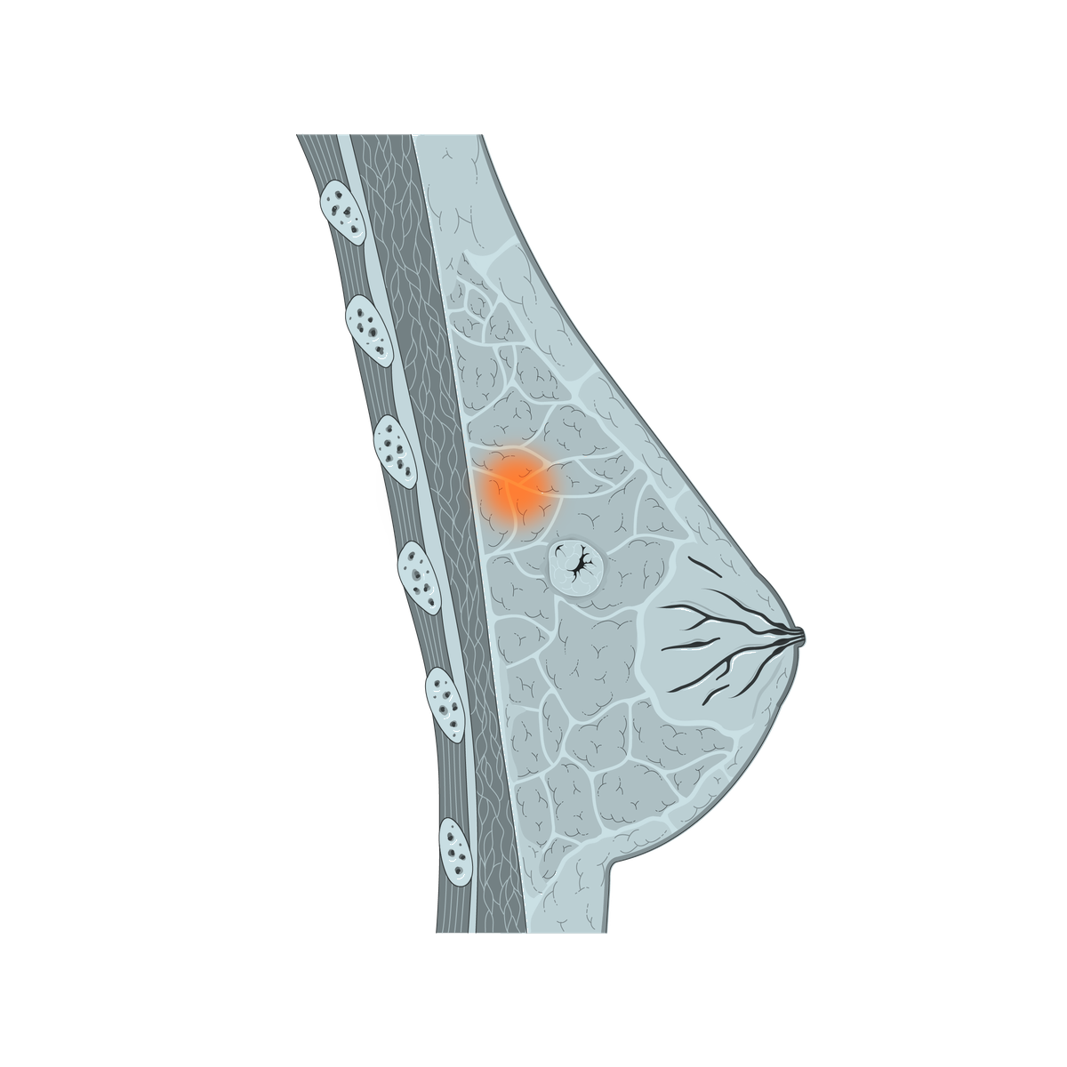
Duct ectasia and nipple discharge
Milk ducts may widen or become inflamed, leading to nipple discharge. The cause should be assessed, and many cases are managed medically with follow-up.
Breast & Oncology Surgery · Korba, Heliopolis
Breast problems may appear as a lump, discharge, inflammation, or a recent change in the nipple or skin. A clear plan begins with examination and appropriate tests, followed by medical treatment, monitoring, or surgery according to the diagnosis.
Understanding breast changes
The breast contains glandular tissue, ducts, fat, connective tissue, blood vessels, and nearby lymph nodes. Symptoms may result from benign, inflammatory, breastfeeding-related, or tumour conditions, so their meaning cannot be judged by touch alone.
Breast tissue includes lobules, ducts, fat, and supporting connective tissue.
Lumps, discharge, and any recent change in the nipple, areola, or breast skin need assessment.
Clinical examination, imaging, and biopsy findings are interpreted together.
Conditions that are not necessarily tumours
Inflammation, milk-duct problems, and breastfeeding-related conditions can sometimes resemble a lump. Examination and appropriate tests determine whether the condition needs medication and monitoring or a limited procedure.
Milk ducts may widen or become inflamed, leading to nipple discharge. The cause should be assessed, and many cases are managed medically with follow-up.
Mastitis or cellulitis of the breast skin may cause pain, redness, and warmth. Treatment often begins with medication and follow-up.
An abscess is a collection of pus that needs assessment and treatment. It may require incision or drainage and cleaning alongside medical treatment.
A milk-filled collection can form during breastfeeding. Assessment determines whether conservative treatment and follow-up are enough or drainage is needed.
The key principle: not every breast problem is a tumour, but persistent symptoms should not be ignored. The diagnosis determines the appropriate treatment.
When might surgery become part of the plan?
Surgery is discussed when removing tissue is expected to clarify the diagnosis, relieve symptoms, or form part of treatment. The recommendation follows the combined clinical, imaging, and pathology findings.
Some benign lesions may be removed when they enlarge, cause discomfort, distort the breast, recur, or remain uncertain after assessment.
Surgical removal may be advised when needle sampling does not provide enough certainty or when the findings remain concerning.
Surgery is commonly part of treatment for operable breast cancer, with the extent tailored to the tumour and the overall treatment plan.
Nearby lymph nodes may be assessed in selected cancer cases to help guide staging and further treatment.
Assessment pathway
The aim is to reach the clearest possible diagnosis before deciding whether monitoring, surgery, or another treatment pathway is appropriate.
Reviewing symptoms, when the change appeared, personal medical history, and family history of breast tumours, followed by examination of both breasts, the nipple and skin, and nearby lymph-node areas.
Ultrasound, mammography, or other imaging may be selected according to age, symptoms, and the clinical findings.
A needle biopsy can provide tissue for microscopic examination and is often essential before planning cancer treatment.
Clinical, imaging, and pathology findings are considered together rather than in isolation.
Monitoring, surgical removal, cancer treatment planning, or referral for additional specialist care as appropriate.
The outcome is not the same for everyone: Many breast changes are benign. Prompt assessment is still important because the correct next step depends on establishing what the change represents.
Age and sex do not rule out a breast condition: Breast conditions and tumours can affect men, although less commonly, and benign or malignant lumps can appear at different ages. A new change should not be dismissed because a patient is young, while recognising that breast-cancer risk generally rises with age.
Surgical options
The operation may range from removing a limited area to removing the whole breast. The choice depends on diagnosis, tumour size and location, breast size, pathology, and the wider treatment plan.
The surgeon determines the appropriate procedure after discussing benefits, alternatives, and the considerations specific to the individual condition.
When surgery is selected
Exact arrangements depend on the operation and the final diagnosis. Instructions are individualized and may involve coordination with radiology, pathology, oncology, or other specialists.
Confirming imaging, biopsy findings, medications, and the planned operation.
Completing preparation and consent according to the hospital plan.
Removing the agreed breast tissue and assessing lymph nodes when indicated.
Monitoring recovery, pain control, the wound, and any drains if used.
Examining the removed tissue to confirm the diagnosis, margins, and other relevant features.
Reviewing healing and pathology, then discussing whether further treatment or surveillance is needed.
Prepare for the consultation
Previous reports and images help create a clearer clinical picture and may avoid unnecessary repetition of some investigations.
Frequently asked questions
This information is general; medical assessment determines what applies to each individual condition.
No. Many breast lumps are benign. However, a new lump or persistent breast change should be assessed so that the cause is identified rather than assumed.
Yes. Breast conditions can affect men, although less commonly, and benign or malignant lumps may appear at different ages. Any new or persistent change should therefore be assessed rather than judged by age or sex alone.
No. Some conditions need only removal of the lump or tumour with a margin. Mastectomy is reserved for situations in which it is considered the more appropriate option.
In selected breast-cancer cases, nearby lymph nodes are assessed to help understand whether cancer cells have spread and to guide the wider treatment plan.
Yes. Surgically removed breast tissue is normally sent for pathology examination. The result can confirm the diagnosis and provide information needed for follow-up treatment planning.
Bring breast imaging, biopsy or pathology results, previous operation reports, a medication list, and any other relevant medical records.
This page provides general information to help patients understand the assessment pathway. It does not diagnose a condition or determine treatment without a medical consultation.
Specialist consultation
You can book a consultation to review breast symptoms, imaging, and biopsy results and discuss whether monitoring, surgical removal, or further specialist treatment planning is appropriate.